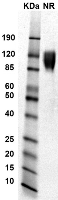
 MW: Molecular Weight marker reduced condition
NR: PD-1 dimer under non-reducing condition
MW: Molecular Weight marker reduced condition
NR: PD-1 dimer under non-reducing condition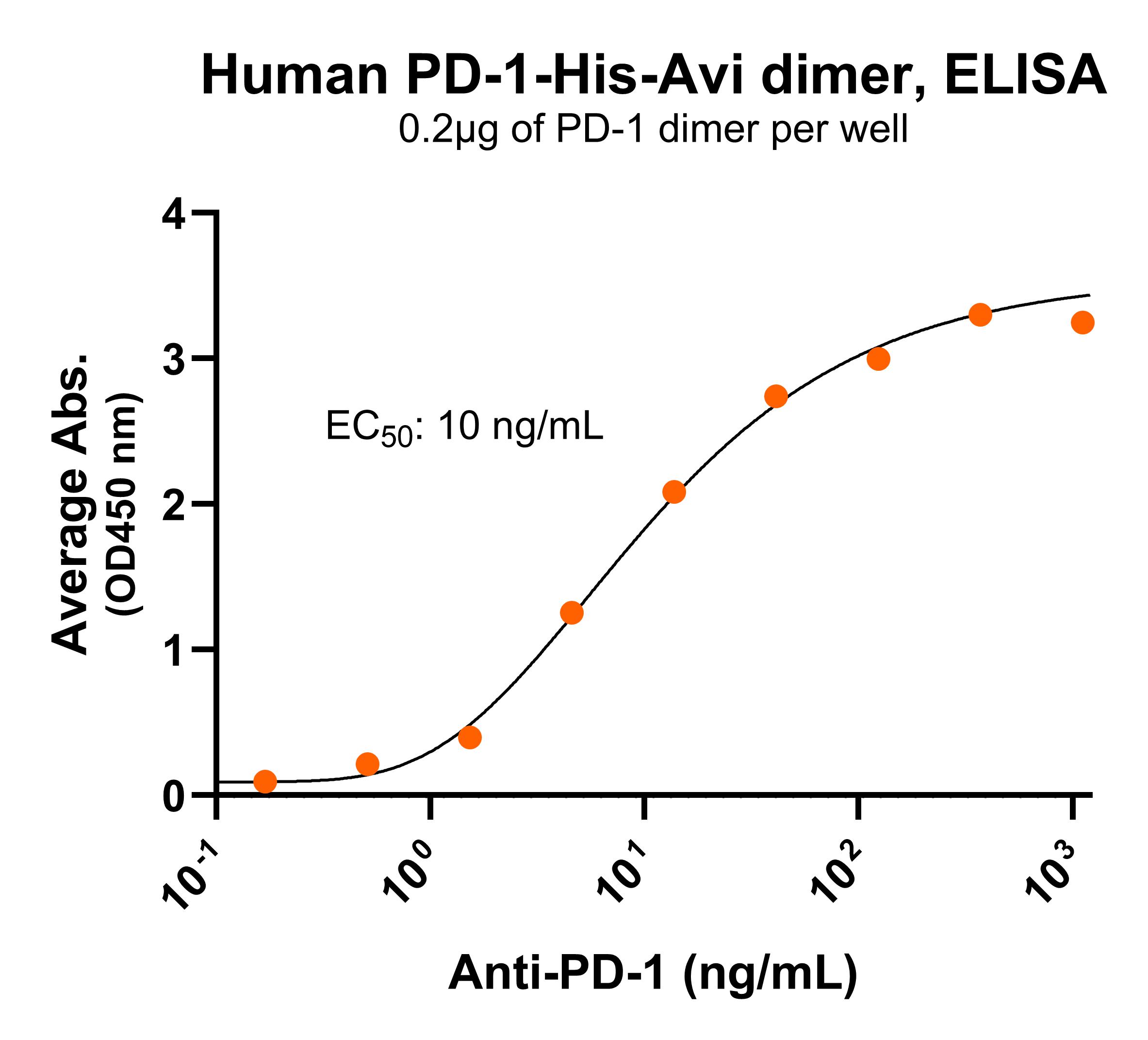 Immobilized human PD-1-His-Avi dimer protein (CSP-24093-03) at 2 μg/mL (100 μL/well) can bind anti-human PD-1 monoclonal antibody with half maximal effective concentration (EC50) range of 5-20 ng/mL (QC tested).
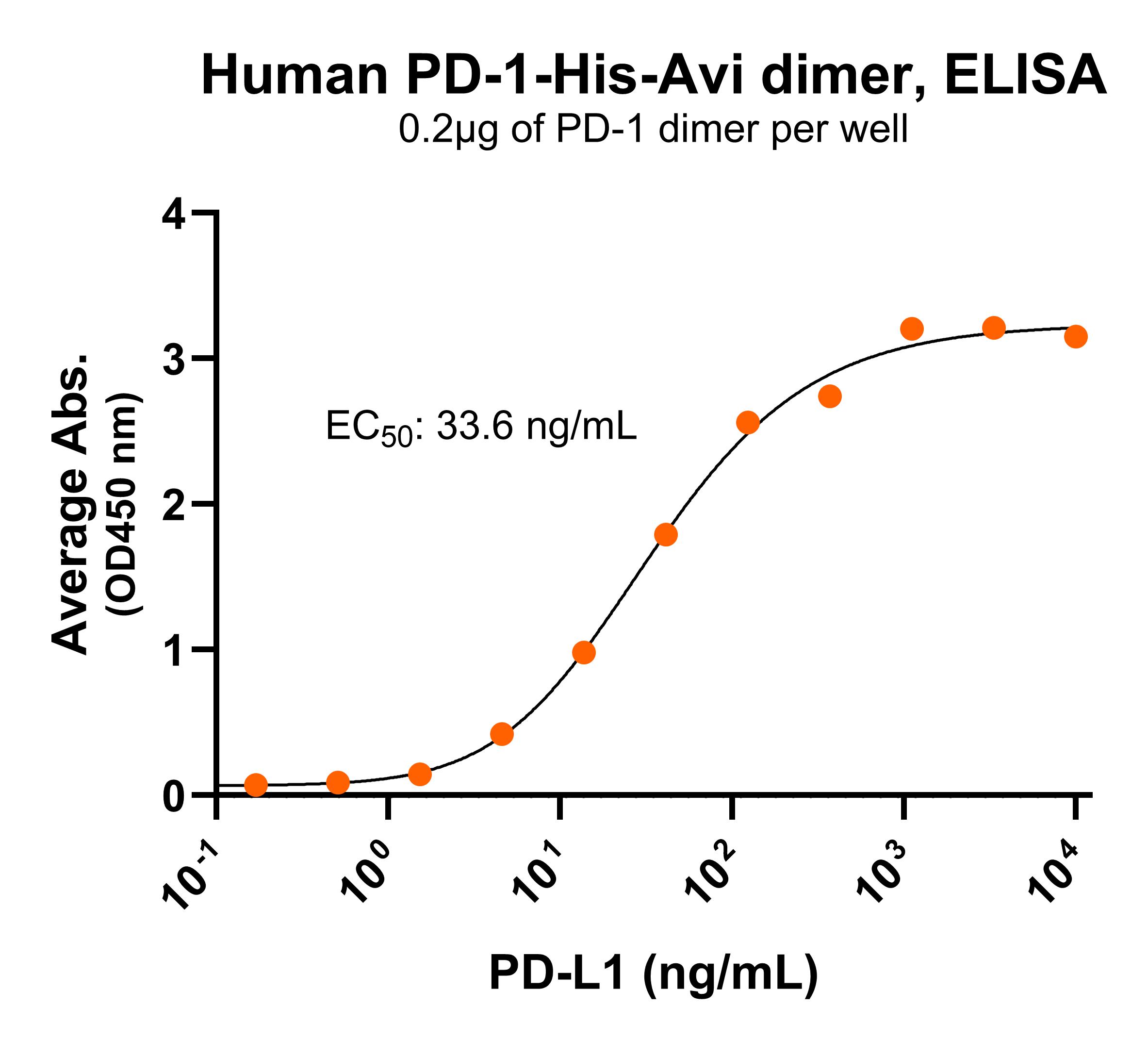 Immobilized human PD-1-His-Avi dimer protein (CSP-24093-03) at 2 μg/mL (100 μL/well) can bind human PD-L1 with half maximal effective concentration (EC50) range of 16.8-67.1 ng/mL (QC tested).